

What is the hip labrum?
The hip labrum is a gasket-like structure that lines the periphery of the acetabulum, or socket of the hip joint. Like the gaskets that seal the joints between the pipes in your house, the acetabular labrum provides a suction seal for your hip joint — adding to the stability to the joint. Similar to rubber, it is made of a spongy substance referred to as fibrocartilage that is susceptible to injury and degeneration.
In addition to providing a stabilizing suction seal for the hip joint, it provides several other key roles in the health of your hip. Those functions include: 1) protective sensation of hip pain referred to as nociception, 2) helping to sense where your hip joint is in space in the coordination of complex lower extremity movements which is referred to as proprioception, as well as 3) dissipating pressure of each of your steps over the entirety of the joint which helps to preserve the health of your cartilage and avoid the development of arthritis.
Given the complexity of the labrum’s roles in the hip joint, the medical literature supports efforts to preserve its presence when tearing or degeneration occur, rather than its removal.
What happens when the labrum tears?
Tears of the labrum can be divided into several different categories that include both acute or sudden traumatic injury or tearing, or chronic conditions leading to degeneration or fraying of the tissue. When disruption of the normal tissue occurs, hip pain and dysfunction often ensue. This manifests as pain in the groin, or front of the hip, mechanical catching or seizing sensations, and pain with deep flexion or twisting at the hip that is common to athletic activities.
Individuals of all ages and activity levels can sustain injuries to the labrum of the hip. Like older pipe gaskets that can dry and crack, aging labral tissue becomes more brittle. Degenerative fraying of the labrum is therefore extremely common in the aging athlete. Studies have shown that approximately 70% of adults will have labral tears identified on MRI studies of the hip, regardless of the presence or absence of pain.
As the presence of hip labral tears is very common, it is critical that symptoms of hip pain are evaluated critically by an experienced hip specialist to help identify why your hip labral tear has occurred and guide your choice of the most appropriate treatment option to fit your desired activity level and goals.
Several factors are important in guiding the treatment of a labral tear. Those factors include the underlying shape of both the ball and socket of the hip. Deep sockets and shallow sockets place extra stress on the labral tissue making it susceptible to tearing. A bony prominence of the edge of the femur, or ball portion of the joint, referred to as a cam deformity can also lead to tearing of the labrum. The shape of the hip joint is best evaluated with special x-rays, which is why they are often obtained as the first step in the evaluation of hip pain. Additionally, the amount of hip joint space narrowing, or arthritis, is also an important factor that is best evaluated with plain x-rays.
Acute twisting or jamming injuries can also lead to shearing of the labral tissue from its boney attachment. Additionally, fraying of the labral tissue naturally occurs in a more chronic fashion with arthritis, or loss of cartilage, of the hip joint. Treatment options vary considerably in all of these scenarios.
How are labral tears treated?
Treatment begins with the appropriate identification of a symptomatic labral tear. After x-rays are obtained, MRI scans provide the image detail necessary to evaluate the labrum and the adjacent cartilage for damage. Treatment of labral tears often begins with a period of relative rest, use of anti-inflammatory medications, a course of physical therapy to strengthen hip and core musculature, and consideration of therapeutic injections.
If these measures fail to get the individual back to their desired activity level, surgical options may be considered. In individuals who lack arthritic changes, arthroscopic surgery – which utilizes minimally invasive portals in the skin to place a small camera and instruments into the joint – can be implemented to fix the labrum. If the socket is too deep or boney prominences are present, the hip can be reshaped to remove the source of impingement to prevent reinjury. In rare circumstances, the labrum cannot be repaired. In those circumstances, either removal or replacement of the labral tissue in the form of reconstruction may be more appropriate options (for more information regarding this new technique see: https://www.ncbi.nlm.nih.gov/pubmed/26524556 ).
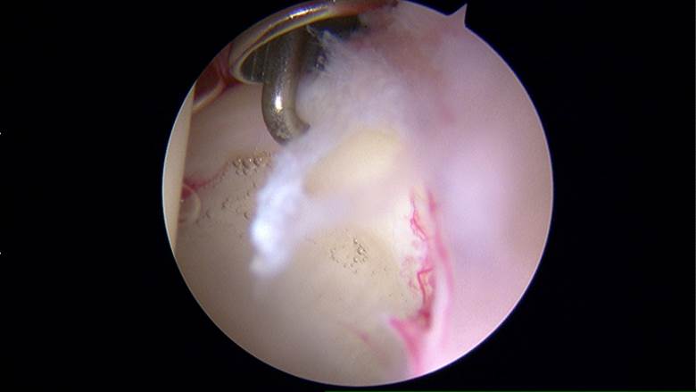
Image 1: An arthroscopic image of a hip labral tear. Frayed tissue is seen in the foreground and a metal probe is demonstrating the separation of the labrum from the rim of the socket.
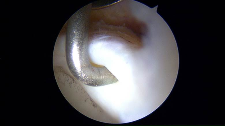
Image 2: A metal probe is further demonstrating separation of the labrum from the rim of the socket.

Image 3: The torn labrum has been repaired with three anchors – securing it back to the rim of the socket.
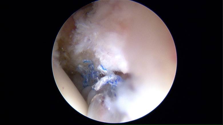
Image 4: The suction seal of the labrum has been reestablished following repair.
Ultimately, it is the goal of the hip specialists at OrthoVirginia to make sense of the available treatment options to help keep you active – regardless of the cause of your hip pain.